Dr. Jérémie BOISSEAU
Specialist in Endodontics
35 B Chemin des Graviers Blancs
FR-25000 – BESANÇON
FRANCE
Clinical case commented by Pr. Jean-Marie Vulcain
Case presentation:
The patient reported severe pain and swelling from the upper right second molar (17). There was tenderness on biting and swelling buccally. The tooth was restored with a metal ceramic crown.
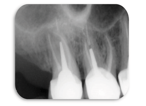
Radiographically it was obvious that the tooth had been previously root treated and that there was:
- Evidence of persistent periapical pathology and bone loss associated with the buccal roots
- The previous root filling appeared to be well condensed and of adequate length
- An inlay-core was present as well as a post within the palatal root
- There is a suggestion of some space beneath the inlay-core, possibly indicating the unsealing of the cement.
Planned re-treatment
- Removal of the crown and the inlay-core
- Inspection and assessment of the remaining tooth tissue under the crown
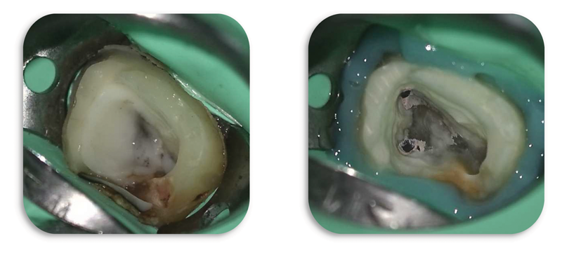
Removal of the remnants of the Glass Ionomer Cement, caries removal and creation of a cervical seal. Initial inspection under the endodontic microscope revealed the presence of a previously untreated MB2 canal. It also became clear that the previous obturation had been completed using a carrier based device, possibly Thermafil.
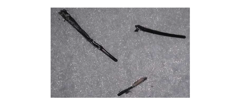
Removal of the carrier based obturation (Thermafil)
- Access cavity preparation
- Ultrasonic preparation uncovering the root canal entries including the MV2 canal
- Use of Neoniti C1 25/.12 and then irrigation
- Root canal patency with Neoniti GPS file, with thorough irrigation at each stage
- Working Length (WL) determination
- Careful shaping of the canal, using the wall brushing technique ensuring that all areas of the canal are prepared including the ones not treated yet with Neoniti A1 no. 20/.06 and A1 no. 25/.06.
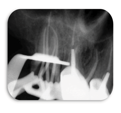
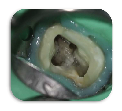
Intraoperative radiography with master cones in place and clinical view of the tooth after root canal preparation
It should be noted the presence of the 4 canals (including the MV2) with their curvature
- Drying of the root canals
- Obturation by warm vertical condensation of the apical third and coronal backfill with injected heated gutta percha. Only the apical half of the palatal canal was obturated, leaving space for the placement of a post
- Each stage of the procedure was performed with the assistance of a surgical microscope.
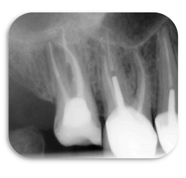
Postoperative radiography of provisional coronal filling
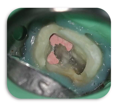
Clinical view after obturation
- The tooth was provisionally restored ensuring a good coronal seal and the definitive restoration of the tooth was scheduled a few days after the endodontic procedure.
Re-treatment of the upper left first molar #26.
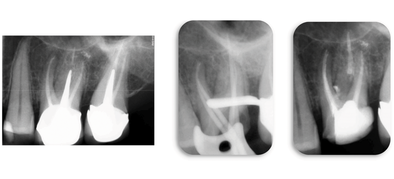
The presence of persistent periapical bone loss associated with the mesial root of the upper right first molar indicates the need for non-surgical re-treatment of this tooth.
The procedure was performed in the same way as Case 1 and the postoperative radiograph shows the presence of a large lateral canal in the coronal third of the mesial root. The treatment of the additional MB2 canal is also evident.
Summary of these cases :
These two cases highlight that endodontic success can only be expected if the treatment is performed with careful adherence to the essential principles of shaping, cleaning, obturation and the provision of a coronal seal. In these cases endodontic retreatment was required. The successful revision of a root treatment, requires a very careful removal of the previous root filling, achieving apical patency and the thorough debridement of the canal space, including preparation of the canal walls using the technique of wall brushing motions to facilitate the thorough irrigation of the root canal system.
The smart use of only 4 Neoniti instruments (C1 no. 25/.12, GPS no. 15/.03, A1 no. 20/.06 and A1 no. 25/.06) resulted in:
- The retreatment of the canals respecting the fundamental principles of endodontics. - Dense three-dimensional obturation by warm vertical condensation. - The subsequent placement of well-sealed and functional coronal restorations.
Ultimately, two teeth that would otherwise have been extracted, were preserved and will serve the patient well for many years.